Imaging Services

When you need imaging services for an accurate diagnosis or advanced treatment, Radiology at Mount Sinai is here for you. We use low-dose imaging with state-of-the-art equipment to provide a full range of radiology services. We have specialists in every area of diagnosis, prevention, and treatment. Our focus is always on you.
Some of these imaging services require special preparation.
Breast Imaging
The Dubin Breast Center is one of the first facilities in the United States to offer state-of-the-art 3D Breast Imaging. This gives us even more information than the standard digital 2D mammography. To obtain a 3D mammogram, we take 15 extremely low dose images of the breast as the mammogram machine rotates in an arc. The computer processes these images and allows us to as 1 mm-thin sections. In this way, we can see small cancers (or healthy tissue) that might otherwise be hidden by overlying tissue.
Computed Tomography
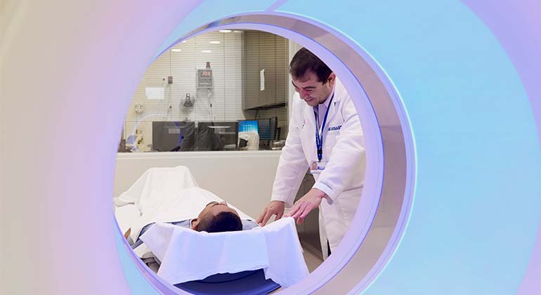
We perform high-resolution computed tomography (CT) scanning of all body areas. CT scans use X-rays and computer software to give doctors pictures of your organs, bones, and other tissues. It helps us see inside your brain, head and neck, spine, and chest, and allows us to examine your cardiovascular, abdominal, pelvic, genitourinary, and musculoskeletal areas. Also called a CAT scan, this test takes 10 to 30 minutes. In addition, we do several types of specialized CT testing:
- CT Angiography. Our 8-, 40-, and 64-slice multi-detector CT scanners focus on blood vessels. They help us get detailed, clear, images very quickly, using minimal radiation. We can even see lesions that are smaller than a centimeter with high clarity. The equipment allows us to use a variety of sophisticated software applications.
- CT Colonography. We use this minimally invasive approach to screen for colonic polyps. Our 8- and 16-slice multi-detector CT scanners produce high-resolution images of the colon quickly. They let us see polyps as small as 1 millimeter.
- CT Coronary Artery Calcium Scoring. This technique helps us see coronary artery calcifications and compare them to national standards. This information enables us to give you a calcium index or “score” that describes your risk for coronary artery disease.
- CT Low-Dose Lung Cancer Screening. When we need to assess your risk of developing lung cancer, we use our 8- and 16-slice multi-detector CT scanners. The scans give us quick and clear images that show nodules smaller than 1 centimeter. We use this approach as part of our Lung Cancer Screening Program.
Dual-Energy X-ray Absorptiometry
This procedure, also called DXA or DEXA, is the most advanced approach to measuring bone density. It is a type of bone mineral density (BMD) exam and tells us if you have osteoporosis. The test measures bone density in the spine and hip.
Emergency Imaging
Our specialists are prepared to handle all emergency imaging needs, 24/7. We read radiological screening on all adult and pediatric emergency room patients anywhere in the Mount Sinai Health System. We interpret X-rays, ultrasounds, computed tomography (CT), and magnetic resonance imaging (MRI) scans. We work closely with all emergency room doctors throughout the health care system.
Magnetic Resonance Imaging
High resolution MRI studies use a large magnet, radio waves, and a computer to show cross-sectional images of your organs and structures. The procedure does not use radiation. MRI scans can take 30 to 45 minutes. We use an MRI to diagnose and treat concerns in the brain, head and neck, spine, chest, cardiovascular, heart, abdomen, pelvis, genitourinary, and musculoskeletal areas. Because we can get the images quickly, these approaches are well suited for children and those uncomfortable in enclosed spaces. We offer several specialized MRI services.
- Functional MRI (fMRI) studies and functional MR imaging of brain activation. Our 1.5 and 3.0 Telsa MRIs quickly provide 3D images of your brain.
- High-yield MRI. This tool allows us to get high quality images of your injury without putting your entire body in a tube. The exams are more comfortable than standard MRI tests. The ONI MSK Extreme system delivers optimal imaging of the hand, wrist, elbow, foot, ankle, and knee.
- Magnetic resonance angiography (MRA). This tool helps us diagnose diseases of the aorta, coronary, carotid, renal, abdominal, and peripheral arteries. These images help us evaluate atherosclerotic disease, congenital heart disease, and cardiac function.
Musculoskeletal Imaging
Being enclosed in a tight space for imaging testing can be uncomfortable. Our advanced MRI technology lets us scan your hand, arm, leg, or foot just by putting your limb inside the device. The technology, the Optima™ MR430s from GE Healthcare, produces very high-quality images.
Positron Emission Tomography
Positron emission tomography (PET) scans use radioactive dye to produce 3D images of your tissues and organs. These scans help us see any changes in your cell function or disease. We often use PET scans together with CT or MRI scans. PET/CT imaging combines unique functional analysis with anatomical precision and the right low dose for each individual patient while still maintaining excellent image quality. PET scans are especially useful in evaluating a single lung nodule; lung or colon cancer; melanoma; esophageal cancer; lymphoma; head and neck cancer; and staging of breast cancer as well as prostate cancer. We also use PET scans to learn about refractory (or uncontrolled) seizures, myocardial viability, and dementia and Alzheimer's disease.
Ultrasound
Ultrasound imaging uses high-frequency sound waves to produce pictures of the inside of the body. This procedure helps us see how your internal organs are functioning and how blood flows through the blood vessels. Ultrasound does not use ionizing radiation (X-rays). We use ultrasound to diagnose conditions and guide biopsies. Most ultrasound examinations are painless, fast, and easy. You lay on the examination table. Then we apply a warm gel on your skin and move a hand-held device called a transducer back and forth over the area we are observing until we get the images we need. If this area is tender, you might feel pressure from the transducer. Ultrasound exams in which the transducer is inserted into an opening of the body (such as transvaginal exams) may produce minimal discomfort. Usually, this exam takes about 30 minutes.
X-Ray
X-rays create images of tissues and structures in the body. We use X-rays to diagnose conditions in the chest, musculoskeletal, abdominal, gastrointestinal and genitourinary X-ray studies. We also perform X-rays of the joints (called arthrography) and spine (myelography).
Treatment
In addition to helping with diagnosis, our specialists also perform treatment procedures. We treat a variety of conditions, from liver and kidney disease to peripheral artery disease to benign prostate hyperplasia (BPH). Our two main approaches are:
- Our interventional radiology experts specialize in minimally invasive, targeted treatments). We move tiny instruments such as catheters (small tubes) through blood vessels to treat various conditions non-surgically.
- Nuclear medicine offers innovative treatment through pills, injections, and inhaled medicines.
Why Mount Sinai?
All imaging at Mount Sinai is supervised and interpreted by a team of specialists with many years of experience. You and your doctors benefit from the expertise of faculty who continue to contribute to the body of world knowledge in imaging through research and innovation. Our faculty regularly lectures at national and international conferences.