Nephrocalcinosis
Nephrocalcinosis is a disorder in which there is too much calcium deposited in the kidneys. It is common in premature babies.

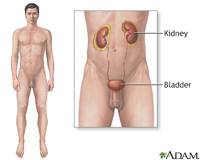
The urinary system is made up of the kidneys, ureters, urethra and bladder.
Causes
Any disorder that leads to high levels of calcium in the blood or urine may lead to nephrocalcinosis. In this disorder, calcium deposits in the kidney tissue itself. Most of the time, both kidneys are affected.
Nephrocalcinosis is related to, but not the same as, kidney stones (nephrolithiasis).
Conditions that can cause nephrocalcinosis include:
- Alport syndrome
- Bartter syndrome
- Chronic glomerulonephritis
- Familial hypomagnesemia
- Medullary sponge kidney
- Primary hyperoxaluria
- Renal transplant rejection
- Renal tubular acidosis (RTA)
- Renal cortical necrosis
Other possible causes of nephrocalcinosis include:
- Ethylene glycol toxicity
- Hypercalcemia (excess calcium in the blood) due to hyperparathyroidism or other medical conditions
- Hypercalciuria (excess calcium in the urine)
- Use of certain medicines, such as acetazolamide, amphotericin B, and triamterene
- Sarcoidosis
- Tuberculosis of the kidney and infections related to AIDS
- Vitamin D toxicity
Symptoms
Most of the time, there are no early symptoms of nephrocalcinosis beyond those of the condition causing the problem.
People who also have kidney stones may have:
- Blood in the urine
- Fever and chills
- Nausea and vomiting
- Severe pain in the belly area, sides of the back (flank), groin, or testicles
Later symptoms related to nephrocalcinosis may be associated with long-term (chronic) kidney failure.
Exams and Tests
Nephrocalcinosis may be discovered when symptoms of renal insufficiency, kidney failure, obstructive uropathy, or urinary tract stones develop.
Imaging tests can help diagnose this condition. Tests that may be done include:
- Abdominal CT scan
- Ultrasound of the kidney
Other tests that may be done to diagnose and determine the severity of associated disorders include:
- Blood tests to check levels of calcium, phosphate, uric acid, and parathyroid hormone
- Urinalysis to see crystals and check for red blood cells
- 24-hour urine collection to measure acidity and levels of calcium, sodium, uric acid, oxalate, and citrate
Treatment
The goal of treatment is to reduce symptoms, prevent more calcium from building up in the kidneys, and reduce kidney damage.
Treatment will involve methods to reduce abnormal levels of calcium, phosphate, and oxalate in the blood and urine. Options include making changes in your diet and taking medicines and supplements.
If you take medicine that causes calcium loss, your health care provider will tell you to stop taking it. Never stop taking any medicine before talking to your provider.
Other symptoms, including kidney stones, should be treated as appropriate.
Outlook (Prognosis)
What to expect depends on the complications and cause of the disorder.
Proper treatment may help prevent further deposits in the kidneys. In most cases, there is no way to remove deposits that have already formed. Many deposits of calcium in the kidneys do not always mean severe damage to the kidneys.
Possible Complications
Complications may include:
- Acute kidney failure
- Long-term (chronic) kidney failure
- Kidney stones
- Obstructive uropathy (acute or chronic, unilateral or bilateral)
When to Contact a Medical Professional
Contact your provider if you know you have a disorder that causes high levels of calcium in your blood and urine. Also call if you develop symptoms of nephrocalcinosis.
Prevention
Prompt treatment of disorders that lead to nephrocalcinosis, including RTA, may help prevent it from developing. Drinking plenty of water to keep the kidneys flushed and draining will help prevent or decrease stone formation as well.
References
Bushinsky DA. Kidney stones. In: Melmed S, Auchus RJ, Goldfine AB, Koenig RJ, Rosen CJ, eds. Williams Textbook of Endocrinology. 14th ed. Philadelphia, PA: Elsevier; 2020:chap 32.
Chen W, Bushinsky DA. Nephrolithiasis and nephrocalcinosis. In: Johnson RJ, Floege J, Tonelli M, eds. Comprehensive Clinical Nephrology. 7th ed. Philadelphia, PA: Elsevier; 2024:chap 60.
Tublin M, Levine D, Thurston W, Wilson SR. The kidney and urinary tract. In: Rumack CM, Levine D, eds. Diagnostic Ultrasound. 5th ed. Philadelphia, PA: Elsevier; 2018:chap 9.
Vogt BA, Springel T. The kidney and urinary tract of the neonate. In: Martin RJ, Fanaroff AA, Walsh MC, eds. Fanaroff and Martin's Neonatal-Perinatal Medicine. 11th ed. Philadelphia, PA: Elsevier; 2020:chap 93.
Version Info
Last reviewed on: 7/1/2023
Reviewed by: Kelly L. Stratton, MD, FACS, Associate Professor, Department of Urology, University of Oklahoma Health Sciences Center, Oklahoma City, OK. Also reviewed by David C. Dugdale, MD, Medical Director, Brenda Conaway, Editorial Director, and the A.D.A.M. Editorial team.
