Neck x-ray
X-ray - neck; Cervical spine x-ray; Lateral neck x-ray
A neck x-ray is an imaging test to look at the cervical vertebrae. These are the 7 bones of the spine in the neck.

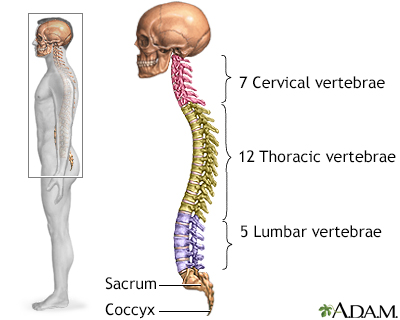
The spine is divided into several sections. The cervical vertebrae make up the neck. The thoracic vertebrae comprise the chest section and have ribs attached. The lumbar vertebrae are the remaining vertebrae below the last thoracic bone and the top of the sacrum. The sacral vertebrae are caged within the bones of the pelvis, and the coccyx represents the terminal vertebrae or vestigial tail.

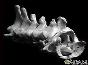
These are the seven bones of the neck, called the cervical vertebra. The top bone, seen on the right of this picture, is called the atlas, and is where the head attaches to the neck. The second bone is called the axis, upon which the head and atlas rotate. The vertebra are numbered from one to seven from the atlas down, and are referred to as C1, C2, C3, etc.

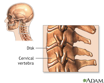
There are seven cervical vertebrae which are located in the neck. They are the smallest, and lightest vertebrae of the vertebral column.
How the Test is Performed
This test is done in a hospital radiology department. It may also be done in your health care provider's office by an x-ray technologist.
You will lie on the x-ray table.
You will be asked to change positions of your neck so that more images can be taken. Usually 2, or up to 7 different images may be needed.
How to Prepare for the Test
Tell your provider if you are or think you may be pregnant. Also tell your provider if you have had surgery or have implants around your neck, jaw, or mouth.
Remove all jewelry.
How the Test will Feel
When the x-rays are taken, there is no discomfort. If the x-rays are done to check for injury, there may be discomfort as your neck is being positioned. Care will be taken to prevent further injury.
Why the Test is Performed
The x-ray is used to evaluate neck injuries and numbness, pain, or weakness that does not go away. A neck x-ray can also be used to help see if air passages are blocked by swelling in the neck or something stuck in the airway.
Other tests, such as MRI, may be used to look for disk or nerve problems.
What Abnormal Results Mean
A neck x-ray can detect:
- Breathing in a foreign object
- Broken bone (fracture)
- Disk problems (disks are the cushion-like tissue that separate the vertebrae)
- Extra bone growths (bone spurs) on the neck bones (for example, due to osteoarthritis)
- Infection that causes swelling of the vocal cords (croup)
- Inflammation of the tissue that covers the windpipe (epiglottitis)
- Problem with the curve of the upper spine, such as kyphosis
- Spine joint that is out of position (dislocation)
- Thinning of the bone (osteoporosis)
- Wearing away of the neck vertebrae or cartilage
- Abnormal development in the spine of a child
Risks
There is low radiation exposure. X-rays are monitored so that the lowest amount of radiation is used to produce the image.
Pregnant women and children are more sensitive to the risks of x-rays.
References
Newton K, Claudius I. Neck trauma. In: Walls RM, ed. Rosen's Emergency Medicine: Concepts and Clinical Practice. 10th ed. Philadelphia, PA: Elsevier; 2023:chap 36.
Sidell DR, Messner AH. Evaluation and management of the pediatric airway. In: Lesperance MM, eds. Cummings Pediatric Otolaryngology. 2nd ed. Philadelphia, PA: Elsevier; 2022:chap 27.
Van Thielen T, van den Hauwe L, Van Goethem JW, Parizel PM. Current status of imaging of the spine and anatomical features. In: Adam A, Dixon AK, Gillard JH, Schaefer-Prokop CM, eds. Grainger & Allison's Diagnostic Radiology. 7th ed. Philadelphia, PA: Elsevier: 2021:chap 47.
Version Info
Last reviewed on: 8/12/2023
Reviewed by: C. Benjamin Ma, MD, Professor, Chief, Sports Medicine and Shoulder Service, UCSF Department of Orthopaedic Surgery, San Francisco, CA. Also reviewed by David C. Dugdale, MD, Medical Director, Brenda Conaway, Editorial Director, and the A.D.A.M. Editorial team.
