Pulmonary ventilation/perfusion scan
V/Q scan; Ventilation/perfusion scan; Lung ventilation/perfusion scan; Pulmonary embolism - V/Q scan; PE- V/Q scan; Blood clot - V/Q scan
A pulmonary ventilation/perfusion scan involves two nuclear scan tests to measure breathing (ventilation) and circulation (perfusion) in all areas of the lungs.
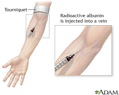
Radioactive albumin injection is part of a nuclear scan test that is performed to measure the supply of blood through the lungs. After the injection, the lungs are scanned to detect the location of the radioactive particles as blood flows through the lungs.
How the Test is Performed
A pulmonary ventilation/perfusion scan is actually 2 tests. They may be done separately or together.
During the perfusion scan, a technician injects radioactive albumin into your vein. You are placed on a movable table that is under the arm of a scanner. The machine scans your lungs as blood flows through them to find the location of the radioactive particles.
During the ventilation scan, you breathe in radioactive gas through a mask while you are sitting or lying on a table under the scanner arm.
How to Prepare for the Test
You do not need to stop eating (fast), be on a special diet, or take any medicines before the test.
A chest x-ray is usually done before, and sometimes after a ventilation and perfusion scan.
You wear a hospital gown or comfortable clothing that does not have metal fasteners.
How the Test will Feel
The table may feel hard or cold. You may feel a sharp prick when the IV is placed in the vein in your arm for the perfusion part of the scan.
The mask used during the ventilation scan may make you feel nervous about being in a small space (claustrophobia). You must lie still during the scan.
The radioisotope injection usually does not cause discomfort.
Why the Test is Performed
The ventilation scan is used to see how well air moves and blood flows through the lungs. The perfusion scan measures the blood supply through the lungs.
A ventilation and perfusion scan is most often done to detect an acute pulmonary embolus (blood clot in the lungs). It is also used to:
- Detect abnormal circulation (shunts) in the blood vessels of the lungs (pulmonary vessels)
- Detect abnormal circulation from multiple old blood clots (chronic thromboembolic disease)
- Test regional (different lung areas) lung function in people with advanced pulmonary disease, such as chronic obstructive pulmonary disease (COPD)
Normal Results
A radiologist will evaluate your ventilation and perfusion scan in conjunction with a chest x-ray. All parts of both lungs should take up the radioisotope evenly.
What Abnormal Results Mean
If the lungs take up lower than normal amounts of radioisotope during a ventilation or perfusion scan, it may be due to any of the following:
- Airway obstruction
- COPD
- Pneumonia
- Narrowing of the pulmonary artery
- Inflammation of the lungs due to breathing in a foreign substance (pneumonitis)
- Pulmonary embolus
- Chronic thromboembolic pulmonary disease
- Reduced breathing and ventilation ability
Risks
Risks are about the same as for x-rays (radiation) and needle pricks.
No radiation is released from the scanner. Instead, it detects radiation and converts it into an image.
There is a small exposure to radiation from the radioisotope. The radioisotopes used during scans are short-lived. All of the radiation leaves the body in a few days. However, as with any radiation exposure, caution is advised for pregnant or breastfeeding women.
There is a slight risk for infection or bleeding at the site where the needle is inserted. The risk with perfusion scan is the same as with inserting an intravenous needle for any other purpose.
In rare cases, a person may develop an allergy to the radioisotope. This may include a serious anaphylactic reaction.
Considerations
A pulmonary ventilation and perfusion scan may be a lower-risk alternative to pulmonary angiography for evaluating disorders of the lung blood supply.
This test may not provide a definite diagnosis, particularly in people with lung disease. Other tests may be needed to confirm or check for the findings of a pulmonary ventilation and perfusion scan.
This test has largely been replaced by CT pulmonary angiography for diagnosing pulmonary embolism. However, people with kidney problems or an allergy to contrast dye can more safely have this test.
References
Herring W. Nuclear medicine: understanding the principles and recognizing the basics. In: Herring W, ed. Learning Radiology: Recognizing the Basics. 5th ed. Philadelphia, PA: Elsevier; 2024:e Appendix e1-e20.
Morris TA, Rose A. Pulmonary thromboembolism: presentation and diagnosis. In: Broaddus VC, Ernst JD, King TE, et al, eds. Murray and Nadel's Textbook of Respiratory Medicine. 7th ed. Philadelphia, PA: Elsevier; 2022:chap 81.
Nair A, Barnett JL, Semple TR. Current status of thoracic imaging. In: Adam A, Dixon AK, Gillard JH, Schaefer-Prokop CM, eds. Grainger & Allison's Diagnostic Radiology. 7th ed. Philadelphia, PA: Elsevier; 2021:chap 1.
Version Info
Last reviewed on: 8/19/2024
Reviewed by: Allen J. Blaivas, DO, Division of Pulmonary, Critical Care, and Sleep Medicine, VA New Jersey Health Care System, Clinical Assistant Professor, Rutgers New Jersey Medical School, East Orange, NJ. Review provided by VeriMed Healthcare Network. Also reviewed by David C. Dugdale, MD, Medical Director, Brenda Conaway, Editorial Director, and the A.D.A.M. Editorial team.
