Protein electrophoresis - serum
SPEP
This lab test measures the types of protein in the fluid (serum) part of a blood sample. This fluid is called serum.

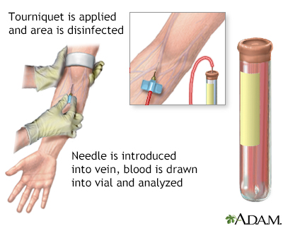
Blood is drawn from a vein (venipuncture), usually from the inside of the elbow or the back of the hand. A needle is inserted into the vein, and the blood is collected in an air-tight vial or a syringe. Preparation may vary depending on the specific test.
How the Test is Performed
A blood sample is needed.
In the lab, the technician places the blood sample on special paper and applies an electric current. The proteins move on the paper and form bands that show the amount of each protein.
How to Prepare for the Test
You may be asked to not eat or drink for 12 hours before this test.
Certain medicines may affect the results of this test. Your health care provider will tell you if you need to stop taking any medicines. Do not stop any medicine before talking to your provider.
How the Test will Feel
When the needle is inserted to draw blood, some people feel moderate pain. Others feel only a prick or stinging. Afterward, there may be some throbbing or a slight bruise. This soon goes away.
Why the Test is Performed
Proteins are made from amino acids and are important parts of all cells and tissues. There are many different kinds of proteins in the body, and they have many different functions. Examples of proteins include enzymes, certain hormones, hemoglobin, low-density lipoprotein (LDL, or bad cholesterol), and others.
Serum proteins are classified as albumin or globulins. Albumin is the most abundant protein in the serum. It carries many small molecules. It is also important for keeping fluid from leaking out from the blood vessels into the tissues.
Globulins are divided into alpha-1, alpha-2, beta, and gamma globulins. In general, alpha and gamma globulin protein levels increase when there is inflammation in the body.
Lipoprotein electrophoresis determines the amount of proteins made up of protein and fat, called lipoproteins (such as LDL cholesterol).
Normal Results
Normal value ranges are:
- Total protein: 6.4 to 8.3 grams per deciliter (g/dL) or 64 to 83 grams per liter (g/L)
- Albumin: 3.5 to 5.0 g/dL or 35 to 50 g/L
- Alpha-1 globulin: 0.1 to 0.3 g/dL or 1 to 3 g/L
- Alpha-2 globulin: 0.6 to 1.0 g/dL or 6 to 10 g/L
- Beta globulin: 0.7 to 1.2 g/dL or 7 to 12 g/L
- Gamma globulin: 0.7 to 1.6 g/dL or 7 to 16 g/L
The examples above are common measurements for results of these tests. Normal value ranges may vary slightly among different laboratories. Some labs use different measurements or test different samples. Talk to your provider about the meaning of your specific results.
What Abnormal Results Mean
Decreased total protein may be due to:
- Abnormal loss of protein from the digestive tract or the inability of the digestive tract to absorb proteins (protein-losing enteropathy)
- Malnutrition
- Kidney disorder called nephrotic syndrome
- Scarring of the liver and poor liver function (cirrhosis)
Increased alpha-1 globulin proteins may be due to:
- Acute inflammatory disease
- Cancer
- Chronic inflammatory disease, such as rheumatoid arthritis and systemic lupus erythematosus (SLE)
Decreased alpha-1 globulin proteins may be a sign of:
Increased alpha-2 globulin proteins may indicate:
- Acute inflammation
- Chronic inflammation
Decreased alpha-2 globulin proteins may indicate:
- Breakdown of red blood cells (hemolysis)
Increased beta globulin proteins may indicate:
- A disorder in which the body has problems breaking down fats (for example, hyperlipoproteinemia, familial hypercholesterolemia)
- Estrogen therapy
Decreased beta globulin proteins may indicate:
- Abnormally low level of LDL cholesterol
- Malnutrition
Increased gamma globulin proteins may indicate:
- Blood cancers, including multiple myeloma, Waldenström macroglobulinemia, lymphomas, and chronic lymphocytic leukemias
- Monoclonal gammopathy of unknown significance (MGUS)
- Chronic inflammatory disease (for example, rheumatoid arthritis, and systemic lupus erythematosus)
- Acute infection
- Chronic liver disease
Risks
There is little risk involved with having your blood taken. Veins and arteries vary in size from one person to another, and from one side of the body to the other. Taking blood from some people may be more difficult than from others.
Other risks associated with having blood drawn are slight, but may include:
References
Lu SX, Rustad EH, Usmani SZ, Landgren CO. Multiple myeloma. In: Hoffman R, Benz EJ, Silberstein LE, et al, eds. Hematology: Basic Principles and Practice. 8th ed. Philadelphia, PA: Elsevier; 2023:chap 91.
Pincus MR, Bluth MH, Brandt-Rauf PW, Bowne WB, LaDoulis C. Oncoproteins and early tumor detection. In: McPherson RA, Pincus MR, eds. Henry's Clinical Diagnosis and Management by Laboratory Methods. 24th ed. Philadelphia, PA: Elsevier; 2022:chap 77.
Rajkumar SV, Dispenzieri A. Multiple myeloma and related disorders. In: Niederhuber JE, Armitage JO, Kastan MB, Doroshow JH, Tepper JE, eds. Abeloff's Clinical Oncology. 6th ed. Philadelphia, PA: Elsevier; 2020:chap 101.
Warner EA, Herold AH. Interpreting laboratory tests. In: Rakel RE, Rakel DP, eds. Textbook of Family Medicine. 9th ed. Philadelphia, PA: Elsevier; 2016:chap 14.
Version Info
Last reviewed on: 3/31/2024
Reviewed by: Todd Gersten, MD, Hematology/Oncology, Florida Cancer Specialists & Research Institute, Wellington, FL. Review provided by VeriMed Healthcare Network. Also reviewed by David C. Dugdale, MD, Medical Director, Brenda Conaway, Editorial Director, and the A.D.A.M. Editorial team.
